Saturday, March 31, 2007

Low Level Laser in Hearing Impairment


From my clinic:

Mrs Mulyani visited my clinic, with her husband, who sufferred hearing difficulty.


Her husband, 39 years old, conducted right ear mastoidectomy on 2003 . Three months ago, left ear worsened. They brought recommendation from an ENT specialist.

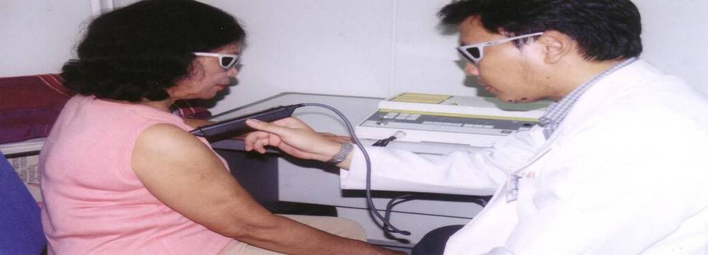
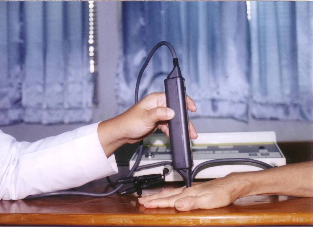
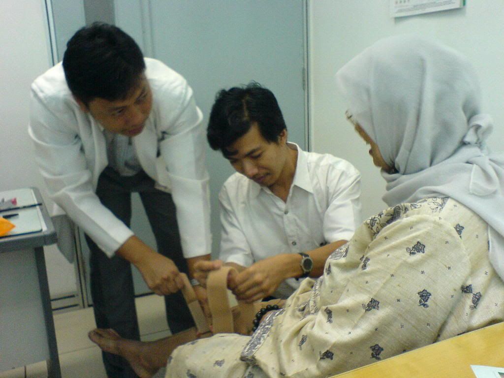
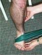
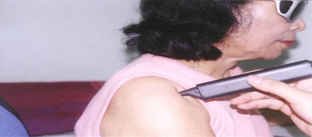
He complaint headache , noisy both ear, poor balance while walking. Low power laser given by ILIB and ear approach, ones a week, for 10 times. ILIB with opening 50 %, for 1 hour, and ear with 650 nm Low laser , 10 mW, 1 hour, pulse, for both ear. Probe approach given ones, at 9th visit, for neck spasm.


After 4th visite , could hear his son crying and adzan from mosque. .

After 5th, pain killer minimize , muscle relaxant stop .

After 6 visite, could speak with normal pitch, balance going better. No headache anymore.

After 10th: No attandane while walking, no drugs for pain, ear noise very
rare,almost disappear.

Program: going to administer 10 times again, ILIB ,and ear.

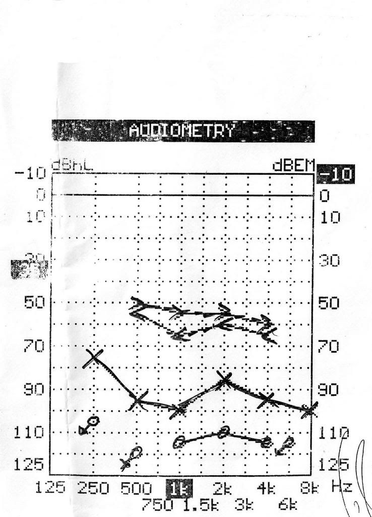
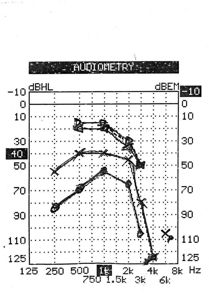
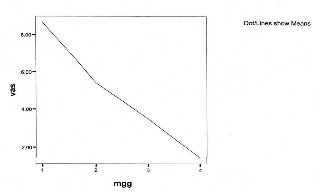
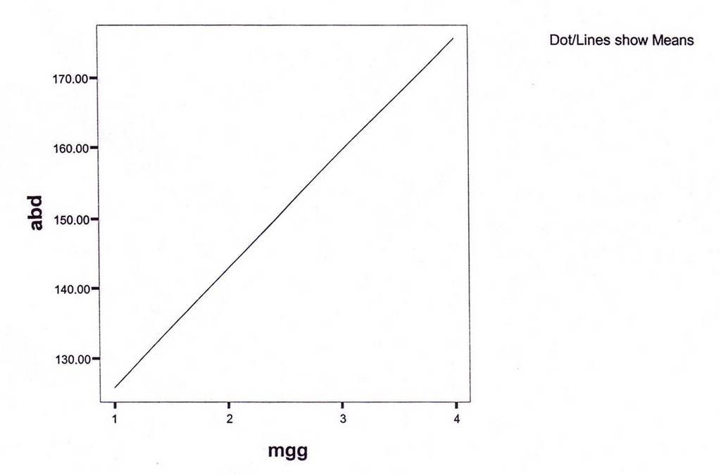
Audimetry: going better compare before LLLT.

(
Before 5 January 2007 - left diagram, After, 20 March 2007 ; right diagram)

Left : April 4, 2006 ; Right : April, 25, 2006

riht ear: increas 39 dB[ left ear increase : 19 dB


Discussion:

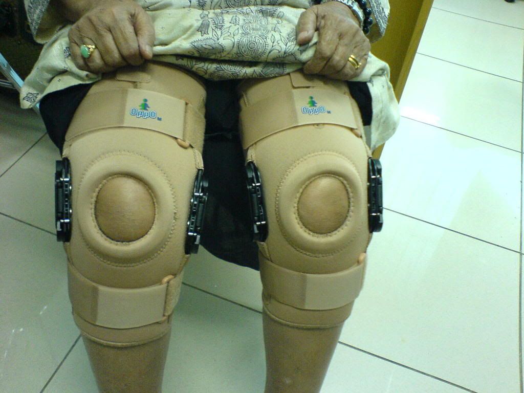
Balance maintance by hearing, visual , and proprioceptive. No doubt he complaint poor balance while walking. To maintain balance, he try to stabilize the neck, so muscles of the neck became stiff.

Balance , and neck muscle pain became better as well as the hearing.

I surprised, he answered my call by cell phone, after 14th therapy.

no pain killer , and muscle relaxant anymore. Tinnitus, gone.

Some journals said , LLLT not work for tinnitus.

May be the dose is to small as well as my experience before I use ILIB , and ear probe.


He is going to work again


From journals:

The low level laser therapy of inner ear diseases has been made use of in Germany since the end of the eighties (for example by Dr. Uwe Witt, Hamburg) and is also to be found in quite a few other countries (Italy, France, Scandinavia, Switzerland, Hungary, the United States of America, Russia etc.).

Since 1991, about 800 inner ear patients have been treated with low level laser light under the medical and scientifical supervision of me. The therapy results have been (and still are) continuously documented and analyzed
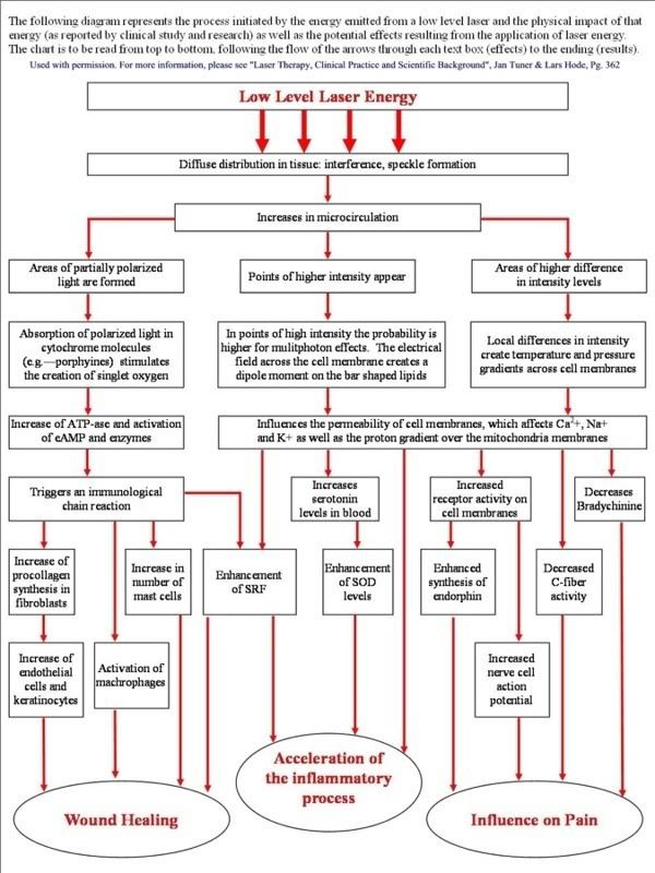
How does LLLT work?

Each cell contains a number of power plants, called mitochondria. The function of these power plants is to produce ATP, the form of energy which can be used by the cell to function properly.

The inner mitochondrial membrane is a large collector surface folded onto a small total volume. All nutritive molecules (fats, proteins and glucose) are oxidized within the cell to the last molecular nutritive component, that is, pyruvate. The pyruvate is then imported into the cellular power plant, where it disintegrates into carbon dioxide and water in the immediate proximity of the collector surface of the mitochondrion. The molecular bond energy thus released, which corresponds with visible, ultraviolet and infrared light as far as its wavelength and frequency is concerned, is utilized by the mitochondrion to produce the cellular fuel ATP.

The collector surface of our cellular power plant is enlarged by the mushroom-shaped protrusions of the inner mitochondrial membrane. The molecular bond energy of the pyruvate is released in the form of light energy. The antennae pigments are capable of absorbing this light energy and transmit it to our cellular power plant, which can now produce the cellular fuel ATP. The natural solar radiation also stimulates the antennae pigments of the mitochondrion to produce ATP.

The cellular power plants can thus produce more ATP. The cellular energy is the fuel the inner ear cells strives after and needs. A sufficiently high supply of cellular energy enables our inner ear cell to work under optimum conditions and is the essential prerequisite to ensure a successful self-healing process.

Low level laser light increases the energy output in our cellular power plant.

(For a detailed discussion, see article: Import of Radiation Phenomena of Electrons and Therapeutic Low-Level Laser in Regard to the Mitochondrial Energy Transfer, Journal of Clinical Laser Medicine & Surgery, Volume 16, Number 3, 1998, Mary Ann Liebert, Inc., Pp.159-165 )
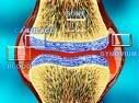
Balance:

Is it possible to help the organ of balance with the high dosage low level lasertherapy by Dr. Wilden¨, specialist of Morbus Menière treatment?
Yes. Vertigo, Morbus Menière and the often in the same time existing sensation of pressure in or surround the ear can be treated very well with the high dosage low level lasertherapy by Dr. Wilden¨, specialist of Morbus Menière treatment.
* from "Das Stato-Akustische Organ" / Reiss-Walkowiak-Zenner-Plinkert-Lehnhardt

Monday, March 19, 2007

Frozen shoulder

Graduated from St Ursula High School - Jakarata. I jointed the millis. I found one of my friends seeks help for her mother. She is suffering couse the pain of her shoulder.I hope this review from one of our research, could help.


Marching brass Putri Santa Ursula


Sonya Monica dearest, this is for you


EFFECTS OF LOW POWER LASER IN FROZEN SHOULDER

Wyasa Andrianto,MD, Ferial Hadipoetro,MD,PhD, Nury Nusdwinuringtyas,MD,

Zuljasri Albar,MD,; Nyoman Murdana,MD, Suryanto Hartono,MD,MA,

Background

Frozen shoulder :

disorder of progressive pain and decrease of range of motion of shoulder articulation

the main causation of shoulder pain and dysfunction in the middle and older age population

treatment option:

ice cooling, medicaments, exercise, TENS, ultrasound diathermy

Objectives

Low power laser therapy with exercise will decrease shoulder pain and increase the range of motion of the shoulder articulation

improve the quality of life of patients with frozen shoulders

Materials and methods

Design comparison of parameter before and after therapy of frozen shoulder patients

OPD Rehab.Med.,Rheumatology Div,Dept.Internal Med,School of Medicine,University of Indonesia/Dr.Cipto Mangunkusumo Hospital,Jakarta,Indonesia

December 2004 to April 2005

INCLUSION CRITERIA

Frozen shoulder patients not less than 18 years of age

VAS not less than 8

Cooperative

Informed consent

Exclusion criteria

Cases with trauma of shoulder articulation

Rheumatoid arthritis and spondyloarthritis with shoulder involvement

Following or under corticosteroid injection therapy within the last 3 months

Patients with abduction range of motion less than 90 degree

patients with contraindication of laser therapy

patients who are unable to perform shoulder exercise properly


Laser device

Endolaser 476(Enraf-Nonius)

Wavelength : 830 nm

Output : 30 mW

Dose : 30 J per treatment

Continuous wave

3 times per week for 3 weeks


DATA ANALYSIS

Data of patients’ background were analyzed by description and were presented in the form of frequency distribution.

Data of VAS and shoulder ROM before and after treatment were analyzed by t test in normal data distribution and were analyzed by Wilcoxon test when data distribution were abnormal.


Table 1.

Group distribution of frozen shoulders

by age


Age(year)

Total

Percentage

40 – 50

51 – 60

61 – 70

1

2

13

6,25

12,5

81,25


Table 2.

Group distribution of frozen shoulders by sex

Sex

Total

Percentarge

Male

Female

6

7

46,15

53,85


Table 3.
Group distribution of frozen shoulders

by education level

Education level

Total

Percentage

Elementary School

Junior High School

Senior High School

Nursing School

College

University

3

1

3

2

3

4

18,75

6,25

18,75

12,5

18,75

25

Table 4.
GROUP DISTRIBUTION OF

FROZEN SHOULDER BY PROFESSION

PROFESSION

TOTAL

PERCENTAGE

PRIVATE

HOUSEWIFE

RETIRED/JOBLESS

NURSE

TEACHER

4

6

4

1

1

25

37,5

25

6,25

6,25

Table 5.
GROUP DISTRIBUTION OF FROZEN SHOULDERS

ACCORDING TO DURATION OF SICKNESS

DURATION

TOTAL

PERCENTAGE

<>

³ 2 months

3

13

18,75

81,25

Table 6.
GROUP DISTRIBUTION OF

FROZEN SHOULDERS BY LOCATION

Location affected

Total

Percentage

Left shoulder

Right shoulder

9

7

56,25

43,75

Table7.
VAS BEFORE AND AFTER TREATMENT OF

FROZEN SHOULDER

VAS SCORE

MEAN± SD

n = 16

p

VAS (pre)

VAS (post)

8,6875 ± 0,8237

1,4125 ± 0,9142


0,000


Graphic 1.

Graphic : Declining VAS score (shoulder joints)


Graphic 2.

Graphic : Increasing joint movements (shoulder joints)


RESULTS AND DISCUSSION

Significant decrement of shoulder pain before and after treatments following 3 weeks of therapy

Significant increment of range of motion of shoulder articulations before and after treatments of low power laser therapy with shoulder exercises following 3 weeks of therapy

CONCLUSIONS

LOW POWER LASER THERAPY AND EXERCISES OF SHOULDER ARTICULATION IS EFFECTIVE AND EFFICIENT MODALITIES TO TREAT FROZEN SHOULDER


Friday, March 16, 2007

Rheumatoid Arthritis


Pingkan's story


Pingkan is in hospital now. She hospitalized due to pain on her both hands and both knees. The working diagnosis is suspect Rheumatoid Arthritis (RA) . The pain very annonying her. I promised to explain to her about RA. but I also ask her to write the history about her illness.
While waiting her story,below a highlight about RA

What is Rheumatoid arthritis (RA)

Rheumatoid arthritis is a chronic, inflammatory, multisystem, autoimmune disorder. It is commonly polyarticular, i.e. it affects many joints. The joints are usually affected initially asymmetrically and then in a symmetrical fashion as the disease progresses. The pain generally improves with use of the affected joints, and there is usually stiffness of all joints in the morning that lasts over 1 hour. The pain of rheumatoid arthritis is usually worse in the morning.

Dear Pingkan, how about your pain?

Deformities

As the pathology progresses the inflammatory activity leads to erosion and destruction of the joint surface, which impairs their range of movement and leads to deformity. The fingers are typically deviated towards the little finger (ulnar deviation) and can assume unnatural shapes. Classical deformities in rheumatoid arthritis are the Boutonniere deformity (Hyperflexion at the proximal interphalangeal joint with hyperextension at the distal interphalangeal joint), swan neck deformity (Hyperextension at the proximal interphalangeal joint, hyperflexion at the distal interphalangeal joint). The thumb may develop a "Z-Thumb" deformity with fixed flexion and subluxation at the metacarpophalangeal joint, and hyperextension at the IP joint.


basic rehabilitation treatment principles
are:
1. relieve pain
2. prevent joint damage and eformities
3. maintain strength and function
4. educate the patient and family
5. help the patient adapt emotionally to life-style limitations
imposed by the diseases proses.

Lovely Pingkan. May be we should try to use Low Laser for you to relive pain.
Would you please click my blog about: Low Level laser in Rheumatoid Arthritis:
the paper just won the 3rd place free paper competition !

(Conclusion : Low level LASER therapy combine with isometric hand strengthening exercise can reduce pain and increase the MCP range of motions in patients with hand rheumatoid arthritis )


It is safe for you?

Phototherapy is FDA approved for a number of applications and has been deemed safe. It also requires relatively little time to perform. Established protocols and tissue dosages have been established that make clinical application relatively easy

Why you dear? Let's look the epidemiology

Epidemiology

The incidence of RA is in the region of 3 cases per 10,000 population per annum. Onset is uncommon under the age of 15 and from then on the incidence rises with age until the age of 80. The prevalence rate is 1%, with women affected three to five times as often as men. It is 4 times more common in smokers than non-smokers. Some Native American groups have higher prevalence rates (5-6%) and black persons from the Caribbean region have lower prevalence rates. First-degree relatives prevalence rate is 2-3% and disease concordance in monozygotic twins is approximately 15-20%


Saturday, March 10, 2007

Philosophy of Rehabilitation Medicine


World Health Organization (1980)


q Impairment : any loss or abnormality of psychological, physiological, or anatomical structure or function

q Disability : any restriction or lack resulting from an impairment of the ability to perform an activity in the manner or within the range considered normal for human being

q Handicap : a disadvantage for a given individual , resulting from an impairment or a disability, that limits or prevents the fulfillment of the role that is normal for that individual


Conventional Medical Model Versus Rehabilitation Model

The general orientation of the medical model is toward disease, while rehabilitation medicine is toward disability, or more broadly , illness.

Diseases is defined as the interaction of a pathologic process with individual molecules , cells, and organs. It is essentially a biological event.

Disability or illness, is essentially a human event. It represents the resulting interaction of a person with a disease.


Physician’s Role

In the medical model, the physician role tends to be active. It is a physician who does the examination, orders the tests, makes a diagnosis, abd prescribes appropriate medications. The physician role in rehabilitation model also encompasses these functions but extends to include helping the patient adjust to the disability and problem solving to minimize the functional loss from a long-term, chronic condition.

Patient’s Role

In the medical model, the patient’s role is often passive and uninformed , with diagnosis and therapeutic measures done or given to him. By contrast , in the rehabilitation model, the patient is encouraged to be an active, informed participant.

Friday, March 9, 2007

OSTEOARTHROSIS GENU- REHABILITATION MEDICINE APPROACH

Impairment, Disability, and Handicap


Other terms associated with rehabilitation include impairment, disability, and handicap. Impairment is the residual limitation resulting from disease, injury, or a congenital defect. Disability is the inability to perform some key life function.
Handicap is the interaction of a disability with the environment.

Report from my office

Mrs. Emma was sent to me with working diagnosis: bilateral knee osteoarthritis. Using rehabilitation terminology, knee osteoarthrosis (OA genu) was her impairment. No limitation to perform activity of daily living (eating, bathing, grooming, toileting), means no disability. Chief complaint: very painful when walking (Visual Analog Scale/ VAS > 6), Handicap : yes.

Program : Goal : pain reduction

Regiment: Diode laser, output 90mW; 60 J , 3 times, every other day

Knee support

Discussion : Low Level Laser given to reduce pain.

Another modality, such as TENS, would not be helpful if VAS for pain > 6

Knee support : to reduce weight bearing , pain reducing is the result

Resulted: mobility: minimally pain while walking after 3rd cold laser ;

no more handicap

Rehabilitation working diagnosis: Impairment : yes :OA Genu

Disability : no

Handicap : no

Program : Impairment: - glucosamine-chondroitine; Quadriiceps isometric exrecise


Knee support for Mrs. Emma - bilateral


Knee support check out


Smiling happily !!!


ANKLE SPRAINS - REHABILITATION


FROM : SKYLARK MEDICAL CLINIC

Ankle Sprains

Ankle sprains are one of the most common sports medicine injuries.
The ankle is stabilized by 3 ligaments on the outside - the anterior talofibular, calconeal fibular and posterior talofibular ligaments (see figure-1st panel, and by a fan-like ligament in the inside-the deltoid ligaments)

Inversion injuries (turning the ankle inward, strain the outer ligaments, while eversion injuries will sprain the inner deltoid ligament. Sprains are classified by degree of severity.

1st degree sprains involve stretching of the ligaments but not tears. There is minimal swelling and instability. Usually the patient may resume sports within a few weeks. This is most common in ankle injury.
2nd degree injuries involve tearing of ligaments with more swelling and bruising. This may take 3-8 weeks to return to sports.
3rd degree injuries involve severe tears of the ligament (yet surgery is rarely required). Because the ligaments are torn it may take 8-12 weeks to heal. Laxity and instability can result of this injury. Many injuries with incomplete tears fall between 2nd and 3rd degree tears.

Treatment of Ankle Sprains
The first thing to do is accurately diagnosing an ankle sprain and not missing other serious fractures that require other treatment.Any injury that is very painful or unable to walk on should be suspected as a fracture and be seen by a doctor. The severity of the injury should dictate the treatment. Even a severe ankle sprain should be kept splinted and on crutches (even if no fracture is seen on the x-ray) since this treatment will also help them heal


Approach to Rehabilitation
Stage 1
- Inflammation (3 days) It is important to rest and splint the ankle. PRICES or Protection, Rest, Ice, Compression, Elevation and Support, will help decrease the pain and swelling associated with the acute injury. An anti-inflammatory drug may be used as well. That will treat both the inflammation and the pain.
Stage 2 - Early mobilization and strengthening (up to 1week) This involves walking on injury (assisted with crutches for partial support if needed). Taping or a brace will also give support. Early exercises to maintain range of motion are important.
Stage 3 - Rehabilitation This involves improving ankle strength, flexibility, and balance. Physiotherapy or athletic therapy is important (this may begin on the 2nd week)
Stage 4 - Late Rehabilitation It is important to ensure that the ankle is adequately strong enough to allow return to sport.


Some advocate 'over-rehabilitation' which refers to the continuing of stretching and exercise programs at home to ensure that this same injury will not reoccur. Often making the athlete stronger than they were before the injury.

Forms of Treatment

Crutches - should be used whenever the injury is very painful to walk on. Gradually crutches may be used less as the injury heals, by allowing more weight to rest on the foot.


Athletic Taping - supportive athletic tape is applied to the ankle to give support. The tape must be applied by an experienced therapist and not left on overnight as it can interfere with circulation

Ankle Braces - commercial over-the-counter braces give similar support to taping but may be easily applied by the patient and are reusable. They may also be used continually to prevent future ankle injuries.

Ankle braces such as "aircast" or equivalent are useful in limiting the injured ankle motion to an up and down plane (dorsiflexion/plantar flexion) but preventing any rolling over. This allows a quicker return to normal activities without re-injuring the injured ankle.

Medications
As mentioned earlier, anti-inflammatories are used at the onset to control inflammation and pain. They are also of benefit later during rehabilitation.
Analgesics such as Tylenol (acetaminophen) give pain control but not ant-inflammation. Glucosamine is a herbal agent that although slower in onset, has anti-inflammatory effects.
Corticosteroid injections are sometimes used in chronic injuries that still have significant pain and swelling but are not used for fresh injuries.

Tuesday, March 6, 2007

Efficacy of different therapy regimens of low-power laser in painful osteoarthritis of the knee: a double blind and randomized-controlled trial

Efficacy of different therapy regimens of low-power laser in painful osteoarthritis of the knee: a double blind and randomized-controlled trial

Gur A; Cosut A; Sarac AJ; Cevic R; Nas K; Uvar A

Physical Medicine and Rehabilitation ; School of Medicine , Dicle University, Diyarbakir , Turkey.

alig@dicle.edu.tr

BACKGROUND MEDICINE OBJECTIVES: A prospective , double blind, randomized, and controlled trial was conducted in patients with knee osteoarthritis (OA) to evaluate the efficacy of infrared low-power Gallium-Arsenide (Ga-As) laser therapy (LPLT) and compared two different laser therapy regimens.

STUDY DESIGN/ MATERIALS AND METHODS: Ninety patients were randomly assigned to three treatment groups by one of the nontreating authors by drawing 1 of 90 envelops labeled ‘A’ (Group I : actual LPLT consisted of 5 minutes, 3 J total dose +exercise ; 30 patients ), ‘B’ (Group II: actual LPLT consisted of 3 minutes , 2 J total dose + exercise ; 30 patients); and ‘C’ (Group III: placebo laser group + exercise ; 30 patients). All patients received a total of 10 treatments , and exercise therapy program was continued during study (14 weeks). Subjects, physician, and data analysis was complete. All patients were evaluated with respect teek 0o pain, degree of active knee flexion, duration of morning stiffness, painless walking distance and duration , and the Western Ontario and Mc Master University osteoarthritis Index (WOMAC) at week 0,6,1, and 14.

RESULTS: Statistically significant improvements were indicated in respect to all parameters such as pain , function, and Quality of Life (QoL), measures in the post-therapy period compared to pre-therapy in both active laser groups (p< style=""> and in parameters such as pain, and WOMAC of the Group II , were more statistically significant when compared with placebo laser group (p<>

CONCLUSIONS: Our study demonstrated that applications of LPLT in different dose and duration have not affected results and both therapy regiment were a safe and effective method in treatment of knee OA.Copyright 2003 Wiley- Liss, Inc.

PMID: 14677160 (PubMed – indexed for MEDLINE)

Sunday, March 4, 2007

ANKLE SPRAINS

This week I share two cases of mine. Ankle sprains and osteoartritis genu.


Story from my clinic


Mrs Dianiati a lady pulmonologyst from a famous hospital in JakartaIndonesia , slipped her ankle while went around a big mall in south Jakarta. Mrs Dianiati did not realized that her left was not already touch the floor, she felt with her left ankle twist to the middle. It is a classic story about ankle sprain. The owner of the groceries do not understand how important to make different color between different high of the floor.

It was so painful, she could not rise immediately. For few minutes she seated on the floor , her lower extremity, below the knee, began edema suddenly.

Roentgen took the day after (Tuesday), bony intack, so the diagnose was ankle sprain. I asked her to visit my clinic, laser administered, diode laser 90 mW; 12 J/cm2 , with probe mode , totally 10 points.

I met her on Wednesday at Cipto Mangunkusumo, edema was gone, just swollen around her ankle. Mobility independently, used sandals, one crutch as cane, but the pain still annoying. Saturday afternoon (3rd March 2007), I gave her diode again but this time for her pain.

(I wrote this report Sunday, 4th oh March 2007.)

Discussion : At her first visited , the laser given to relive the edema, and second visit for the pain.

Ussually the recovery will take four weeks, but at the first week, she already ambulation with minimal dependent. Edema decrease because effect emitted energy laser to the microcirculation, and influenced on pain due to effect to the C-fiber activity and bradychinine. (I enclose diagram represent the proces initiated by the energy emitted). The dose and the mode the laser delivery take important place to get best result. It is a therapeutic window must be understand, and also the mode to reach the site of the damage.

Low-level laser therapy in ankle sprains as randomized clinical trial

de Bie RA; de Vet HC; Lensen TF; van den Wildenberg FA; Kootstra G; Knipschild PG

Departement of Epidemiology , Mastrich University , The Netherlands

OBJECTIVE : The test the efficacy of low-level laser therapy on lateral ankle sprains as an adition to standardized treatment regiment, a trial was conducted in which high laser (5 J/ cm2), low-dose laser (0.5/cm2),and placebo laser therapy (0J/cm2) att skin laser level were compared .

DESIGN: Randomized , double-blind, controlled clinical trial with a follow-up of 1 year. Patients, therapist, assessors, and analyst were blinded to the assigned treatment.

SETTING: An ambulatory care setting.

PATIENTS: After informed consent and verification of exclusion criteria, 217 patients with acute lateral ankle srains were randomized to three groups from September 1, 1993 , through Desember 31, 1995.

INERVENTIOS: Twelve treatments of 904 nm laser therapy in 4 weeks as an adjunct to a standardized treatment regiment of 4 weeks of brace therapy combined ith standardized home exercise and edvice. The laser therapy device used was a Ga- As laser , with 25-watt peak power and 5,0000 or 500 Hz frequency, a pulse duration of 200nsec, and an irradiated area of 1cm2.

PRIMARY OUTCOME MEASURES : Pain and function as reported by the patient

RESULTS: Intervention –to- treat analysis of the short term results showed no statistically significant difference on the primary outcome measure, pain (p=.02) and at some points for hindrancein activities in daily life and pressere pain, as well as subjective recovery (p+.05). intention-to-treat analysis showed that total days absenteeism from work and sports were remarkably lower in the placebo group than in the laser groups, rangimg from 3.7 to 5.3 and 5 to 8 days, respectively. The total number of relapsesat 1 year in the low-dose laser group (n=22) was significantly higher (p=.04) than in the other two groups (high laser, n=13; placebo, n=13). Subgroup analysis to correct for possible confounders did not alter these findings.

CONCLUSIONS: Neither high- nor low dose laser therapy is effective in the treatment of lateral ankle sprains.

Arch Phys Med Rehabil. 1998 Nov;79(11):1415-20